Loculated Pleural Effusion Cxr : Consolidation and collapse in the right lung with a large ... - Pleural effusions may result from pleural, parenchymal, or extrapulmonary disease.
Loculated Pleural Effusion Cxr : Consolidation and collapse in the right lung with a large ... - Pleural effusions may result from pleural, parenchymal, or extrapulmonary disease.. Most malignant effusions can be controlled by thoracentesis and/or closed thoracostomy tube drainage and sclerosis of the pleural cavity. Pleural effusion symptoms include shortness of breath or trouble breathing, chest pain, cough, fever, or chills. The pleura is a thin membrane that lines the surface of your lungs and the inside of your chest wall. Pleural fluid/serum protein ratio >0.5. Other causes are complicated parapneumonic effusion.
Loculated pleural effusion on cxr. Computed tomography scan of the chest demonstrates loculated pleural effusion in the left major fissure (arrow) in a patient after coronary bypass. Not respond to chest tube and antibiotics. Approximately 1 million people develop this abnormality each year in the united states. It detects pleural effusions with higher sensitivity and specificity than cxr, and provides valuable information about the size and depth of the pleural effusion, the echogenicity of the fluid, the presence of septated or loculated fluid, pleural thickening and nodularity, and the presence of any.
Chest CT scan showing a loculated right-sided pleural ... from www.researchgate.net Symptomatic loculated malignant pleural effusion treatment. Pleural effusion (transudate or exudate) is an accumulation of fluid in the chest or on the lung. If none is present the fluid is virtually always a transudate. Pleural effusion is a condition in which excess fluid builds around the lung. Pleural fluid/serum ldh ratio >0.6. When you have a pleural effusion, fluid builds up in the space between the layers of your pleura. Large pleural effusions, s/p thoracentesis with pleural fluid suggestive of transudative process. The cardiac silhouette is also obscured.
Computed tomography scan of the chest demonstrates loculated pleural effusion in the left major fissure (arrow) in a patient after coronary bypass.
Symptomatic loculated malignant pleural effusion treatment. Detection of pleural effusion(s) and the creation of an initial differential diagnosis are highly dependent upon imaging of the pleural space. Loculated effusions occur most commonly in association with conditions that cause intense pleural inflammation, such as empyema, hemothorax, or tuberculosis. Most malignant effusions can be controlled by thoracentesis and/or closed thoracostomy tube drainage and sclerosis of the pleural cavity. Published online by cambridge university press: A pleural effusion is accumulation of excessive fluid in the pleural space, the potential space that surrounds each lung. Commonly from congestive heart failure or malignancy. If none is present the fluid is virtually always a transudate. Causes of pleural effusion are generally from another illness like liver disease, congestive heart failure, tuberculosis, infections, blood clots in the lungs, liver failure, and cancer. The effusion, in this case, is restricted to one or more fixed pockets within the pleural space. Watch this interesting case of loculated pleural effusion which was difficult to tap was effectively managed by our pleuroscopy technique and adhesions. Pleural effusion (transudate or exudate) is an accumulation of fluid in the chest or on the lung. Other causes are complicated parapneumonic effusion.
Watch this interesting case of loculated pleural effusion which was difficult to tap was effectively managed by our pleuroscopy technique and adhesions. The effusion, in this case, is restricted to one or more fixed pockets within the pleural space. Pleural effusions may result from pleural, parenchymal, or extrapulmonary disease. Pleural fluid ldh > two thirds of upper limit for serum ldh. Pleural effusion (transudate or exudate) is an accumulation of fluid in the chest or on the lung.
Right Subpulmonic Pleural Effusion images, diagnosis ... from www.vcuthoracicimaging.com Other causes are complicated parapneumonic effusion. Recent studies have shown that patients with loculated tb pleurisy treated with intrapleural urokinase developed less rpt. Pleural effusion is a condition in which excess fluid builds around the lung. Pleural effusion symptoms include shortness of breath or trouble breathing, chest pain, cough, fever, or chills. The pleura is a thin membrane that lines the surface of your lungs and the inside of your chest wall. Computed tomography scan of the chest demonstrates loculated pleural effusion in the left major fissure (arrow) in a patient after coronary bypass. Pleural effusion (fluid in the pleural space). Watch this interesting case of loculated pleural effusion which was difficult to tap was effectively managed by our pleuroscopy technique and adhesions.
Pleural fluid/serum protein ratio >0.5.
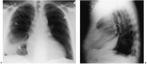
Pleural effusion develops when more fluid enters the pleural space than is removed. More than one half of these massive pleural effusions are caused by malignancy; Large pleural effusions, s/p thoracentesis with pleural fluid suggestive of transudative process. Pleural effusion (fluid in the pleural space). The pleura are thin membranes that line the lungs and the inside of the chest cavity and act to lubricate and facilitate breathing. Most commonly caused by a viral infection. Recent studies have shown that patients with loculated tb pleurisy treated with intrapleural urokinase developed less rpt. Most malignant effusions can be controlled by thoracentesis and/or closed thoracostomy tube drainage and sclerosis of the pleural cavity. When you have a pleural effusion, fluid builds up in the space between the layers of your pleura. The pleura is a thin membrane that lines the surface of your lungs and the inside of your chest wall. A pleural effusion is accumulation of excessive fluid in the pleural space, the potential space that surrounds each lung. Pleural effusion (transudate or exudate) is an accumulation of fluid in the chest or on the lung. There is a large left pleural effusion obscuring the lower half of the left hemi thorax.
Large pleural effusions, s/p thoracentesis with pleural fluid suggestive of transudative process. Always do pleural biopsy if you suspect tb.disorder in the workup of a pleural effusion after performing thoracentesis always order. Loculated effusion (atypical radiological findings). Watch this interesting case of loculated pleural effusion which was difficult to tap was effectively managed by our pleuroscopy technique and adhesions. Most commonly caused by a viral infection.
Disease of the Pleura | Radiology Key from radiologykey.com Most malignant effusions can be controlled by thoracentesis and/or closed thoracostomy tube drainage and sclerosis of the pleural cavity. Excess fluid in the pleural space; The pleura is a thin membrane that lines the surface of your lungs and the inside of your chest wall. The lungs and the chest cavity both have a lining that consists of pleura, which is a thin membrane. Always do pleural biopsy if you suspect tb.disorder in the workup of a pleural effusion after performing thoracentesis always order. Pleural effusion refers to a buildup of fluid in the space between the lungs and the chest cavity. The pleura are thin membranes that line the lungs and the inside of the chest cavity and act to lubricate and facilitate breathing. Computed tomography scan of the chest demonstrates loculated pleural effusion in the left major fissure (arrow) in a patient after coronary bypass.
Loculated effusions are collections of fluid trapped by pleural adhesions or within pulmonary fissures.
An exudative pleural effusion occurs when there is increased permeability of the pleural surface and/or capillaries, usually as a result of inflammation. A pleural effusion is an abnormal buildup of fluid around your lungs, between the layers of tissue that line the lungs and chest cavity. If none is present the fluid is virtually always a transudate. Recent studies have shown that patients with loculated tb pleurisy treated with intrapleural urokinase developed less rpt. Loculated effusion (atypical radiological findings). Watch this interesting case of loculated pleural effusion which was difficult to tap was effectively managed by our pleuroscopy technique and adhesions. Lam s, banim p bmj case rep 2014 apr 9;2014 doi: Pleural effusion (transudate or exudate) is an accumulation of fluid in the chest or on the lung. Pleural effusion (fluid in the pleural space). Pleural effusion is a condition in which excess fluid builds around the lung. The pleura are thin membranes that line the lungs and the inside of the chest cavity and act to lubricate and facilitate breathing. Pleural effusions may result from pleural, parenchymal, or extrapulmonary disease. Most commonly caused by a viral infection.
Pleural effusion symptoms include shortness of breath or trouble breathing, chest pain, cough, fever, or chills loculated pleural effusion. Pleura inflammation, causing sharp pain with breathing;


0 Komentar